

VesselMap
Static Vessel Analysis Software
The unique VesselMap analysis software is used to determine and analyze vascular parameters from previously recorded retinal images. These parameters are
- Risk assessment of vascular-related diseases
- Risk assessment before interventions
- Estimation and prediction of progression
- Testing of therapeutical approaches
Further information: Method of Static Vessel Analysis
- Simple, software-guided application
- Automatic determination of vessel topology with preselection of vessel sections and precise determination of vessel parameters (depending on the fundus camera)
- Integrated function for progress monitoring for a significant improvement in reproducibility and fully automatic evaluation of follow-up images
- Available with German and English user interface
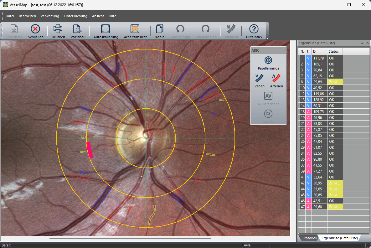
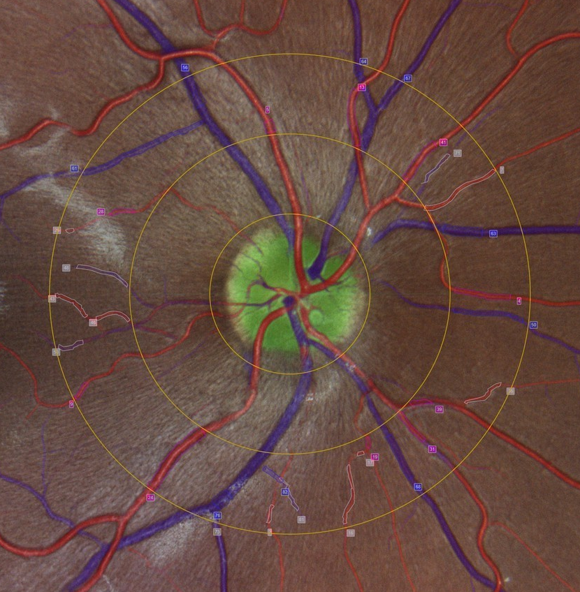
- At least one image of the retina is acquired in a standardized manner with the imaging system
- The evaluation of the digitized fundus image is performed using ring-shaped markers centered to the location of the optic disc
- The software makes an automatic preselection of all detected arterial and venous vessels
- From this, the vessel diameter is automatically averaged spatially-segment-wise for each marked vessel
- In the last step, the vascular parameters are calculated according to the procedures described by Hubbard in the ARIC study [1][2].
[1] Hubbard, Larry D., et al. “Methods for evaluation of retinal microvascular abnormalities associated with hypertension/sclerosis in the Atherosclerosis Risk in Communities Study.” Ophthalmology 106.12 (1999): 2269-2280; doi:10.1016/s0161-6420(99)90525-0
[2] Sharrett A R, et al. “Retinal arteriolar diameters and elevated blood pressure: the Atherosclerosis Risk in Communities Study” Am J Epidemiol 150(3); (1999):263-70; doi:10.1093/oxfordjournals.aje.a009997.
VesselMap determines the following static vessel parameters:
- Central retinal arteriolar equivalent (CRAE): arterial model vessel diameter.
- Central retinal venular equivalent (CRVE): venous model vessel diameter.
- Arteriolar-to-venular ratio (AVR): the CRAE/CRVE ratio.
The central equivalents CRAE and CRVE describe model vessel diameters for characterizing the central vessels. These biomarkers take into account all arterial and venous vessels flowing into and out of the retina according to a geometric-hemodynamic weighting.
VesselMap is available as a software license in combination with terms of 1 year or 10 years. It can be combined with various imaging systems from leading manufacturers and integrated into the existing software environment. The analysis and calculation parameters of the software are tailored to your system and adapted accordingly.
The current VesselMap has not been certified by us as a medical device according to MDR (Medical Device Regulation) due to disproportionately high costs and effort and is only approved for research use. Versions that have already been delivered retain their previous approvals.
In order to integrate the software into your existing environment, a suitable fundus camera and a corresponding computer must be available. We will be happy to check the compatibility of your existing hardware with our products. Alternatively, both can also be obtained from Imedos.
- The imaging unit, for acquiring the fundus images if a suitable system is not yet available.
- The workstation consisting of instrument table, computer unit and power supply for comfortable and mobile work.
- The server solution, for connecting different workstations and synchronizing the corresponding databases.
- The management system connection for smooth integration of static vessel analysis into existing software. (DICOM)
- The research option, for free examination of vessel diameters outside the standardized examination protocol.
