Methods of Retinal Vessel Analysis
Retinal Vessel Analysis, which can be static or dynamic, offers unprecedented opportunities for non-invasive examination of the vital and health-relevant microcirculation. It provides important information about the holistic vascular health of patients. As a “mirror image” of microvascular changes throughout the body, vascular analysis allows important conclusions to be drawn about systemic disease and the development of end-organ damage.
Static vascular analysis - method for non-invasive assessment of vascular condition.
Static Vascular Analysis is an excellent and cost-effective method for non-invasive and non-contact examination of the condition and morphology of microcirculatory vessels. In this examination, retinal arterioles and venules are imaged with a fundus camera to determine clinically relevant parameters of vascular topology. Our extremely precise and easy-to-use camera, developed for this purpose VesselMap analysis software is ideally suited for use in clinical routine. The recorded vascular parameters represent valid biomarkers, confirmed by large studies, which can be used as risk factors or prognostic indicators for vascular diseases and vascular events.
Download:
Product Brochure Static Vessel Analysis (german)
Produkt Brochure Static Vessel Analysis (english)
Dynamic vascular analysis - method for non-invasive examination of microvessel function and autoregulation.

The worldwide unique method of Dynamic Vascular Analysis allows a non-invasive investigation of the functional and autoregulatory mechanisms of the smallest vessels. As an exceptional approach to this microcirculation, vascular analysis at the eye provides essential information on subclinical changes of the entire body. These provide insight into the holistic vascular health of patients and allow important conclusions to be drawn about systemic diseases and the development of end organ damage.
To analyze and quantify these functional autoregulatory mechanisms, our proprietary software measures and evaluates the time-varying vascular topology in video sequences in real time. During these measurements, vascular autoregulatory mechanisms are stimulated, making the resulting dynamic vascular response integral to the analyses.
The Imedos Dynamic Analyzer uses flicker light as a standard functional diagnostic stimulation to study retinal endothelium-dependent microvascular dysfunction. The vascular response is NO-mediated (endothelial NO synthase) and plays a key role among the regulatory mechanisms of vascular autoregulation and in many microcirculatory disorders and vascular diseases of various organs. Dynamic vascular analysis is therefore ideally suited for interdisciplinary use in clinical routine as well as for medical research.
Download:
Product Brochure Dynamic Vessel Analysis (german)
Product Brochure Dynamic Vessel Analysis (english)
Related information online:
Retinal Vessel Analysis: Reflecting the Microvasculature
(Article by R.Gerste in the German Medical Journal)
Endothelial function in cardiovascular medicine
A consensus paper of the European Society of Cardiology Working Groups on Atherosclerosis and Vascular Biology, Aorta and Peripheral Vascular Diseases, Coronary Pathophysiology and Microcirculation, and Thrombosis . 2021 Jan 1;117(1):29-42. doi:10.1093/cvr/cvaa085.
Physical activity and exercise improve retinal microvascular health as a biomarker of cardiovascular risk
A systematic review. Atherosclerosis. 2020 Dec;315:33-42. doi:10.1016/j.atherosclerosis.2020.09.017. Epub 2020 Sep 23. PMID: 33212315.
An aging society and the simultaneous increase in cardio- and cerebrovascular events make timely identification of risk patients and factors desirable. In a variety of incipient systemic diseases, the retinal vessels are affected particularly early. Thus, research and early detection of pathological processes of the retinal circulation are of high importance.
The retina is almost the only place in the human body that allows direct and non-invasive examination of the smallest blood vessels as well as the microcirculation itself. Changes in retinal microcirculation not only have direct effects on the retina and the tissues of the eye, but also allow conclusions to be drawn about systemic changes, especially as biomarkers.
For example, there is an association between retinal changes such as microaneurysms and arteriolar narrowing and the prevalence of coronary heart disease, myocardial infarction, or stroke. (see alsoCooper L. S. et al. 2006; Wong T. Y. et al. 2002; Wong T. Y. et al. 2003])
Retinal arterioles are known to have similar anatomic, physiologic, and autoregulatory properties to those of cerebral and coronary microvessels [Patton N. et al. 2005]. A retinal endothelial dysfunction, which is detected and quantified during Dynamic Vessel Analysis, is considered a strong predictor of long-term cardiovascular events and can be used for risk stratification [ Theuerle J. D. et al. 2021])
In conjunction with various microcirculation-stimulating techniques, phenomena such as neurovascular coupling, Bayliss effect and retinal vascular autoregulation can be visualized, and pathophysiological processes in diseases can be assessed. (see for example [ Hanso et al. 2007])
Fundamentals of the investigation
Our integrated system for dynamic vascular analysis consists of our special fundus camera for imaging in the form of video sequences with our specialized analysis software.
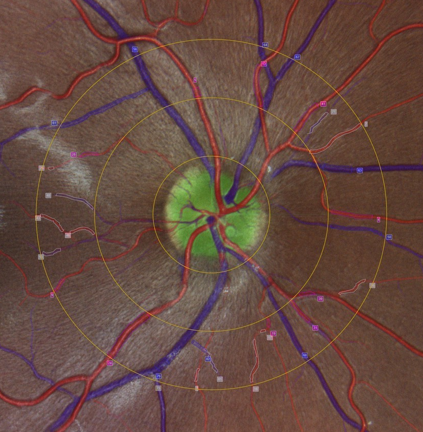
The physiological basis of the examination is the column of red blood cells that forms in the retinal blood vessels and is separated from the vessel walls by the plasma edge current. The red blood cells absorb some of the observation light, allowing the blood vessels to image well on the retina. The proprietary algorithms of our analysis software determine the exact diameter of this column in the image sequences, expressed in measurement units (MU), which reflects the real vessel diameter. For a normal eye according to Gullstrand, 1 MU = 1 µm.
The temporal resolution is 40 ms, i.e. 25 video images per second are
evaluated. This also allows observation of pulsatory effects or phase shifts in the time-varying vessel diameters in relation to other signals, such as the R-wave in the ECG.
Flicker stimulation for the examination of endothelial function
In the course of imaging, the electronics integrated in our analysis system modulate the observation light temporally over the entire 30° field of view of the retinal camera, creating an alternating light-dark contrast.
Stimulation with this flicker light and the accompanying increased neuronal activity induces dilation of retinal vessels. This dilation causes improved blood supply to the retina and optic nerve and is related to the state of microvascular endothelial function and thus the systemic state of the microcirculation. The associated regulatory mechanisms are impaired in a variety of neurodegenerative and vascular diseases [ Garhöfer et al. 2020]) Due to the measurement of the vascular response as a result of flicker stimulation, dynamic vascular analysis provides increased accuracy and can detect even the smallest changes in microcirculation events. The effect of flicker light and the mechanisms of flicker action on the endothelium have been described in the literature, see, e.g., in [ Hanssen et al. 2022])
The chosen frequency of 12.5 Hz of the flicker light provides a sequence of a illuminated and a dark frame at the video frequency of 25 Hz. This frequency is in the range of the maximum exciting flicker frequency. The standard flicker stimulation protocol used consists of a baseline of 50 s with three subsequent flicker periods of 20 s each, each interrupted by 80 s of uniform light as a pause to reset the vascular state. During the study, selected retinal vessel segments are measured in real time and the flicker-induced vessel changes are compared with the baseline topological vessel parameters.
Reference book: "Retinal vessel analysis - a new method of diagnostics and risk prediction"
 The innovative diagnostic approach of Retinal Vessel Analysis is proving to be extremely revolutionary, especially in the interdisciplinary field of preventive medicine. Together with Prof. Dr. Henner Hanssen and eight other scientists, our former managing director Dr. Walthard Vilser, has written the first reference book on this subject. In addition to the methodology, it also presents fields of application, such as ophthalmology and cardiology, as well as current research results.
The work with the ISBN 978-3-9374-1576-6 is published by UNI-MED for 29.80 euros and comprises 63 pages. It can be ordered directly from Imedos Health or from the publisher’s website. The reference book was initially published in English; German and Chinese editions are planned.
