Applications of retinal vessel analysis in research
Vascular analysis procedures are an exceptional opportunity for non-invasive and non-contact examination of the condition and function of the smallest blood vessels in the entire body. The eye as an incomparable access to this microcirculation provides essential information on subclinical changes and provides information on the holistic vascular health of humans. The knowledge gained enables important conclusions to be drawn about systemic diseases and the development of end-organ damage and offers various potential uses in research into future applications in prevention, screening, therapy monitoring and patient motivation.
Retinal Vessel Analysis methods provide important biomarkers about the state and function of retinal vessels and their regulation in the microcirculation. They are an important milestone in research on the way to personalized medicine. Objective personalization is essential here, as vascular function can change completely differently in different people despite having the same BMI or cholesterol levels, for example.
In addition to the important and efficient assessment of cardiovascular risk, the methods of retinal vascular analysis are particularly suitable for:
- The control of therapeutic and preventive measures
- The control of therapy success
- The estimation and prediction of the progression
- Motivating patients to make lifestyle adjustments with appropriate progress monitoring
- The access to the endothelial function
- The development of drugs
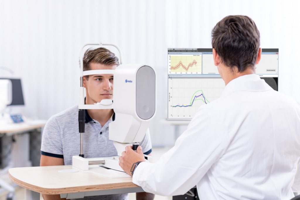
Dynamic vascular analysis with the Imedos Dynamic Analyzer (IDA) can additionally be used for the examination and analysis of retinal endothelium-dependent microvascular dysfunction (MVD). Cardiovascular risk factors such as diabetes, dyslipidemia and arterial hypertension lead to impairment of this vascular function.
Download:
The aim of cardiometabolic prevention is to prevent or at least delay the development and manifestation of vascular changes or diseases at an early stage. This corresponds to the concept of EVA (Early Vascular Aging) and ADAM (Aggressive Decrease of Atherosclerosis Modifiers).
Retinal Vessel Analysis parameters offer new opportunities to extend this concept to include the microcirculation and to place microvascular parameters in the context of established macrovascular gold standards such as flow mediated dilation, intima media thickness, pulse wave velocity and others.
The microvascular risk parameters represent an additional benefit in cardiovascular risk stratification, especially for women. It is desirable to record the retinal microvascular vascular parameters at an early age, as vascular ageing begins very early. Studies show that the first changes in microvascular parameters can already be detected in children using retinal vascular analysis.
The individual influences of lifestyle-changing measures are also reflected in the parameters of the vascular analysis. The parameters of the static vascular analysis are of particular importance, as they can be determined quickly and easily in a non-mydriatic screening. These parameters provide indicators of cardiovascular risk. Studies have shown that lifestyle changes and preventive measures can significantly reduce individual vascular risk. The effect of a measure can be easily observed and is highly motivating for the patient.
From a developmental physiological point of view, the retina with the optic nerve head can at least be regarded as an outsourced part of the brain. There are many similarities and close connections to the brain, not only from the point of view of the optic nerves, but also with regard to microcirculation. It is therefore hardly surprising that studies have established a link between the vascular condition and autoregulatory functions of the retina with depression, Alzheimer’s disease, vascular dementia and stroke.
The parameters of the Static and Dynamic Vessel Analysis allow conclusions to be drawn about the risks of cerebrovascular events such as strokes and the severity of so-called “small vessel diseases”. Changes in retinal vascular analysis parameters are associated with the frequency of white matter lesions on MRI and with the severity of dementia. There is evidence that retinal vascular analysis parameters can also be used to subtype between microvascular and macrovascular strokes.
Dynamic vascular analysis can be used to study not only NO-dependent endothelial function but also neurovascular coupling, myogenic autoregulation and their changes in various systemic diseases such as diabetes mellitus and arterial hypertension and to obtain important information about brain physiology and pathophysiology.
Retinal vascular analysis examines static and dynamic vascular parameters that quantify the steady-state vascular condition and the vascular function or regulatory capacity of the microcirculation and are well-validated cardiovascular risk indicators. Systemic changes in the small arteries and veins of the microcirculation and their regulation occur in a similar form in many organs, including the kidneys.
A large number of scientific publications present the associations between retinal vascular changes, arterial hypertension and renal changes. Very good evidence for the informative value of retinal vascular analysis is provided by study results showing that venous dysregulation of the retinal vessels is a predictor of mortality in patients with end-stage chronic renal failure.
The kidney plays an essential role in the development of systemic arterial hypertension. Conversely, arterial hypertension leads to changes in the microcirculation, which can ultimately be examined in the eye as end-organ damage using retinal vascular analysis.
Organs can only function well with an adequate blood supply. This is particularly true for the eye. The blood supply to the eye must be adapted to current needs at all times, for example to compensate for fluctuations in perfusion pressure, to adapt to changing neuronal activities or to keep the temperature at the fundus constant despite fluctuations in the outside temperature. The blood flow at rest and the ability of the vessels to regulate the blood flow are therefore of central importance.
Impaired blood flow is often the cause of many eye diseases or an important co-factor for the occurrence or progression of the eye disease. Imedos technology makes it possible to diagnose these vascular components and then check the influence of a therapy.
Interdisciplinary approach: In many systemic diseases such as diabetes, hypertension, lipid metabolism disorders or rheumatism, the ophthalmologist can, by examining the blood vessels in the eye, provide colleagues from other specialties with crucial information about the general health of the vessels (the vascular health) and about the response to a therapy or prophylaxis, even in the early stages.
Consequences of circulatory disorders for the eye:
-
The relatively rare acute reductions in blood supply lead to infarction, e.g., of the retina or optic disc
-
The much more common chronic inferior perfusion favors severe disease outcomes and in extreme cases, the formation of neovascularizations
-
Fluctuating oxygen supply due to impaired regulation increases local oxidative stress, a central factor in the pathogenesis of many diseases
-
Hypoxia and oxidative stress damage the blood-brain barrier, favoring the formation of edema and hemorrhage
The focus of Imedos technology is on the retinal vessels because:
-
A functioning retina is essential,
-
The vessels of the retina are optically accessible,
-
The activity of the retina can be increased in a controlled manner by flicker light and thus the ability and capacity of the regulation of the blood vessels can be measured,
-
The retinal blood vessels are not autonomously innervated and vascular endothelial cell function can thus be specifically measured.
Why are vascular endothelial cells of particular interest?
Endothelial cells play a decisive role in the regulation of retinal blood flow. A person has several trillion of these endothelial cells. Interestingly, the state of health of these cells in the retina is a very good indicator of the state of health of these cells throughout the body. In cardiovascular and cerebrovascular diseases as well as in primary vascular dysregulation, the vascular endothelial cells are always affected first.
Fields of application (clinical pictures in ophthalmology)
Retinal arterial occlusions
The examination with the VesselMap on the partner eye provides information on the general state of health of the blood vessels and thus on the risk of further vascular occlusions, whether in the eye or in other organs.
Glaucoma
If damage is progressing in a glaucoma patient despite normal or normalized eye pressure, the SGA and examination with the Imedos Dynamic Analyzer (IDA), whether and in what form a vascular disease is present. If both are disturbed, it is usually a problem in the form of arteriosclerosis and its risk factors. If, on the other hand, the static vascular parameters are unremarkable and the examination with the IDA shows a disturbed endothelial function, a treatable primary vascular dysregulation is present.
If the RVP is increased, the blood flow to the optic nerve head is also reduced. In this case, not only the eye pressure but also the RVP must be lowered.
Diabetic retinopathy
Static vessel analysis with our VesselMap software shows the risk of developing diabetic retinopathy at an early stage. If non-proliferative retinopathy is already visible, it indicates the risk for vascular complications, including the development of proliferative retinopathy.
The examination with the Imedos Dynamic Analyzer shows whether and how well the retinal blood flow can still be regulated. Increased retinal venous pressure (RVP) increases hypoxia and thus the formation of cotton wool spots and neovascularization. High RVP also increases transmural pressure and thus contributes to the formation of edema, hard exudates and hemorrhages.
Chorioretinopathia centralis serosa
In CRCS, local dysregulation of the choroidal vessels can be detected with ICG angiography. However, the frequent underlying dysregulation is also indicated by reduced responses in the DVA and an increase in the RVP.
Retinopathia pigmentosa (RP)
Here, the blood supply to the eye is partly secondary and partly primary. If the examination with the DVA shows endothelial dysfunction, this indicates that one component is primary. In most cases, the RVP is also increased. Both can be improved therapeutically.
Perioperative vision loss
Acute vision loss in connection with operations, e.g. after spinal surgery, is rare but serious. A disturbed regulatory ability, visible in the DVA, explains a lack of or insufficient adaptation, e.g. to a special positioning. If the RVP is elevated, this is a further explanation for the event and enables prophylaxis.
Altitude sickness
If the partial pressure of oxygen decreases, some people develop altitude sickness. Those affected usually already have pre-existing reduced responses in the DVA and then increase their RVP more than others at altitude.
Flammer syndrome (FS)
FS is a general predisposition to react differently to stimuli with the blood vessels. The consequences are particularly common in the eye. The RVP is often increased, the response in the DVA is reduced, but the vessel diameters in the SVA are normal. If this constellation is present, the risk of developing an FS-associated disease is greater.


