
- New design for easier operation
- Painless, non-invasive examination
- High reproducibility of analysis results due to high-precision optics and analysis properties
- Patented internal fixation and macular cover for easier fixation and reduction of light exposure
- Standard examination protocol with stimulation of the retina by flicker light
- Measurement resolution of the diameter of the vessels: <1µm
- Time resolution: 40ms
- Field of view 30°
-
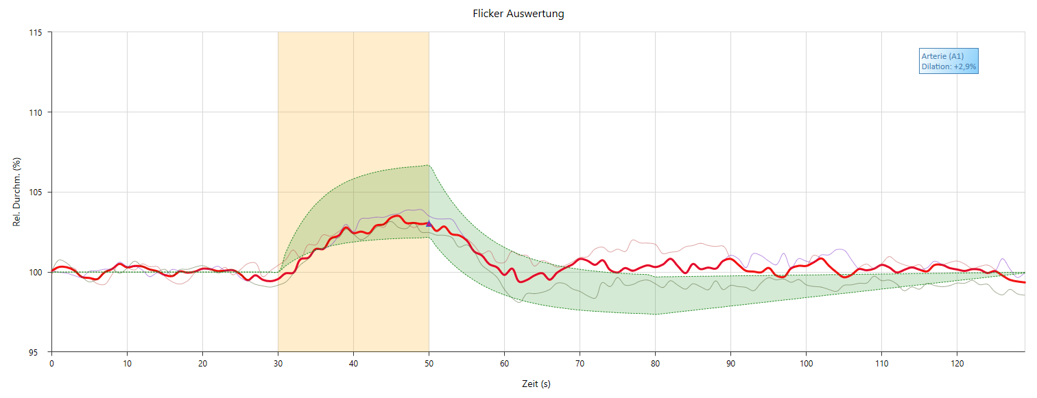
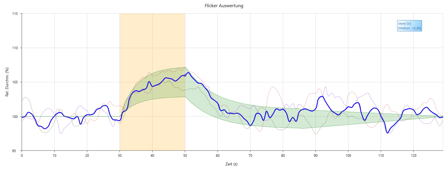
Baseline phase: the baseline state of the retinal vessels is recorded for 50 s to subsequently calculate the dilation or constriction of the vessels in percent to baseline.
-
Stimulation or flicker light phase:For functional diagnostics of the MVD, flicker light is used for 20 s during vessel diameter recording (stimulation phase). The green measuring light is interrupted in the change of the image sequence (12.5 Hz), so that a dark image alternately follows an illuminated one. The recording of the vascular response is continuous and in real time.
-
Post phase: the stimulation phase is followed by a post phase prescribed by the protocol, during which the values of vessel diameters usually return to the baseline level.
-
Repetition: Phases 2 and 3 are repeated twice each.
-
Summary of all phases: The three flicker phases are summarized by signal averaging and graphically displayed as a cumulative curve in the examination result.
The examination protocol of the simple flicker examination is oriented to a strict standardization of the evaluation and is limited to the following three vascular parameters:
- Flicker light induced dilation of the artery (FID art): Arterial dilation maximum of the vascular response to flicker light stimulation in % to baseline.
- Flicker light induced dilation of the vein (FID ven): Venous dilation maximum of the vascular response to flicker light stimulation in % to baseline.
- Flicker light constriction of the artery (FIC art): Arterial constriction maximum of the postphase vascular response to flicker light stimulation in % to baseline.
These vascular parameters characterize the MVD – the function or dysfunction of autoregulation and at the same time the autoregulatory reserve.
Below are sample curves for an arterial and venous flicker response.
- digital video archiving for storage and repeated evaluation of the video files of an examination
- additional measuring points for the simultaneous investigation of up to 10 measuring locations
- modified measurement protocol to adapt the standard protocol to specific medical or medical-experimental issues
- dynamic brightness measurement for the investigation of pulsation phenomena in the capillary bed
The external signal unit is required to connect external devices, such as ECG monitors or blood pressure monitors, to the basic system.
The server solution enables the connection of different workstations and synchronization of the corresponding databases.
TheEvaluation Engine offers versatile possibilities for the individual and grouped evaluation of large amounts of data.
With the purchase of the additional module “Research-Option” you get the possibility to change the standard protocol or even to deactivate the flicker completely. This opens up a multitude of further possibilities with which the various autoregulation or local control mechanisms can be selectively investigated using targeted stimuli.
Some research examples of these include:
- the stimulation by blood pressure elevation to study myogenic autoregulation (Bayliss effect)
- stimulation by inhalation of differently composed respiratory gases, for example 100 percent oxygen, to study the contractility of vascular segments
- the evaluation of pharmacological mechanisms of action
- stimulation with flickering light outside the standard protocol to study neurovascular coupling and vascular endothelial function
- and many more …
